
Case #27
-
History: 54 year old presents with acute left sided weakness.
© 2012 Must See Radiology
History: 54 year old presents with acute left sided weakness.
© 2012 Must See Radiology
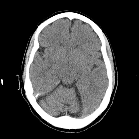
CT head axial, no contrast

CTA head axial, level of MCA
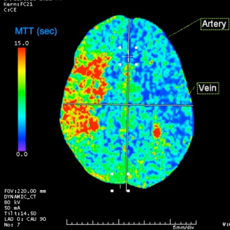
CT Perfusion, Mean Transit Time color image

CT Perfusion, Blood Volume color image

CT Perfusion, Blood Flow color image
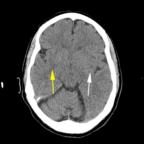
Noncontrast CT head image demonstrates minimal low density change in the right insular ribbon (yellow arrow) compared to the normal left side (white arrow). Finding identified retrospectively.
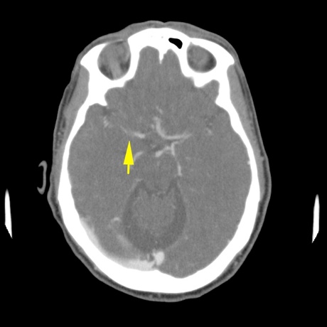
CTA head through the middle cerebral arteries demonstrates discontinuity in the right MCA (yellow arrow) compared to the normal left.
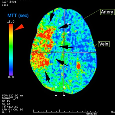
CT Perfusion image demonstrates an area of increased mean transit time in the right frontal lobe, outlined by the black arrows. The left frontal lobe demonstrates normal low mean transit time.
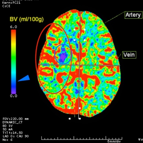
CT Perfusion image demonstrates a small focus of abnormally decreased blood volume in the right frontal lobe, representing an area of infarction. The surrounding area, within the red circle, represents the area of reversible ischemia, the penumbra. *The penumbra is not seen on this image, rather copied over from the Blood Flow images, shown below with the blue circle.
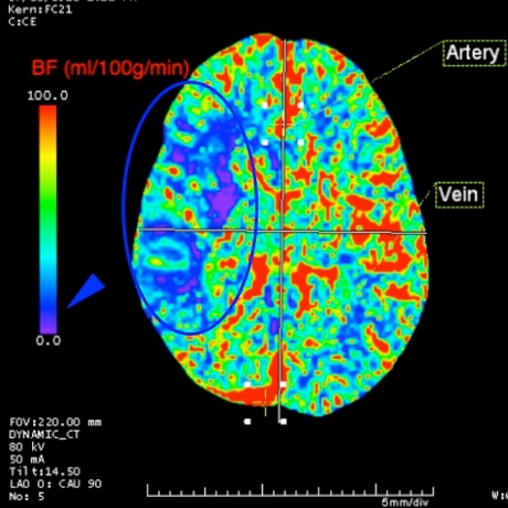
CT Perfusion image demonstrates decreased blood flow in the right frontal lobe (blue circle). This area represents the area of brain ischemia.
KEY_FINDING_3_TEXT
KEY_FINDING_4_TEXT
KEY_FINDING_5_TEXT

KEY_FINDING_6_TEXT

KEY_FINDING_7_TEXT
© 2012 Must See Radiology
Acute right middle cerebral infarction with a large area of reversible ischemia
This case is an example of a patient presenting with acute right MCA stroke. Utilizing the CT perfusion images, we can determine that the patient has a small area of irreversible infarction with a large area of reversible ischemia, detailed in Case Findings.
Additional Information:
Hoeffner, EG. "Cerebral Perfusion CT: Technique and Clinical Applications." Radiology June 2004: 231, 632-644.
© 2012 Must See Radiology
Not available at this time.
Rating not available at this time.
Any feedback regarding this case can be emailed to Tony@mustseeradiology.com
Thank you for trying Must See Radiology!
© 2012 Must See Radiology