
Case #30
-
History: Unconscious patient after traumatic aortic rupture & repair.
© 2012 Must See Radiology
History: Unconscious patient after traumatic aortic rupture & repair.
© 2012 Must See Radiology
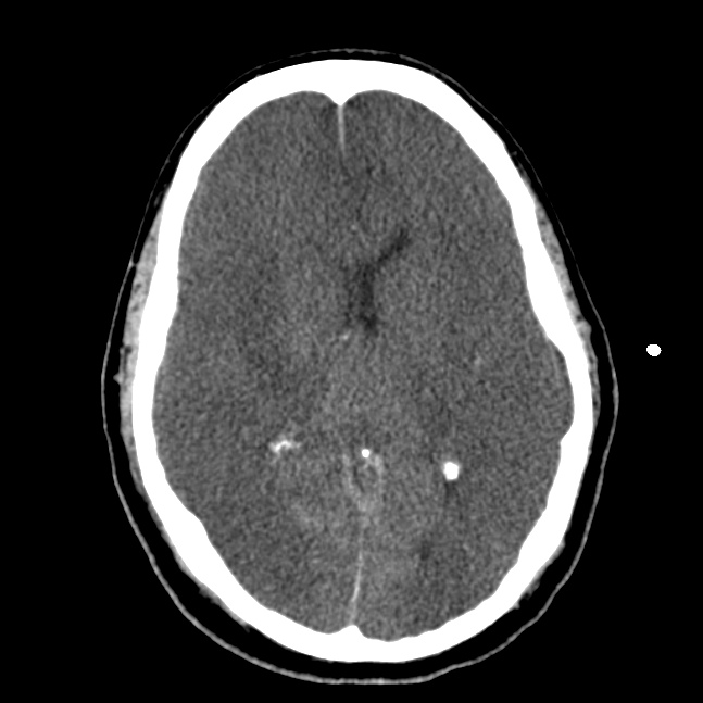
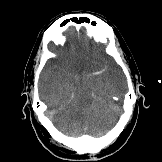
Axial CT images of the head without contrast enhancement
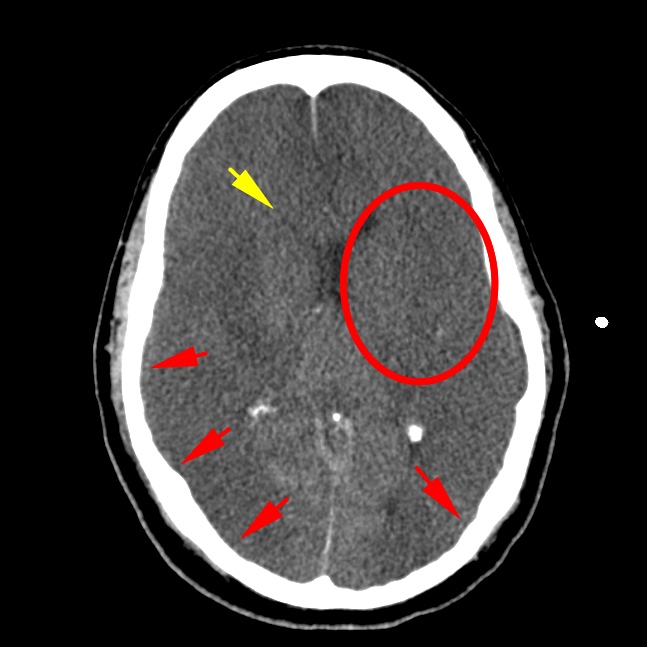
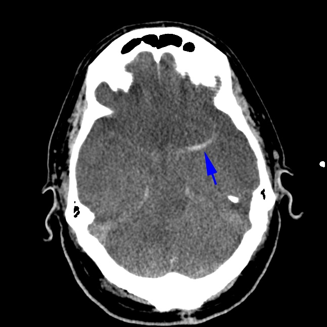
Diffuse low density throughout the cerebral cortex with loss of normal sulci/gyri (red arrows) and grey-white differentiation (red circle). The right frontal horn of the lateral ventricles is slit-like (yellow arrow) due to compression from diffuse cerebral edema. There is a dense left MCA (blue arrow).
© 2012 Must See Radiology
Diffuse Cerebral Edema
due to prolonged traumatic hypoxic event (aortic rupture)
Diffuse Cerebral Edema is a result from severe cerebral ischemia / infarction. Massive brain swelling has a high mortality/morbidity rate.
Signs of Diffuse Cerebral Edema:
- loss / effacement of sulci, especially near the vertex.
- loss of gray-white matter differentiation
- loss of perimesencephalic cistern
Additional Information:
Huang, BY "Hypoxic-Ischemic Brain Injury: Imaging Findings from Birth to Adulthood" Radiographics 2008: 28, 417-439.
Kavanagn, EO "The Reversal Sign" Radiology 2007: 245, 914-915.
© 2012 Must See Radiology
Not available at this time.
Rating not available at this time.
Any feedback regarding this case can be emailed to Tony@mustseeradiology.com
Thank you for trying Must See Radiology!
© 2012 Must See Radiology