MSR

© 2012 Must See Radiology

MSR

Case #31

Key Image

Axial and Coronal contrast enhanced CT images of the chest at the level of the ascending aorta and aortic arch.


Key Findings

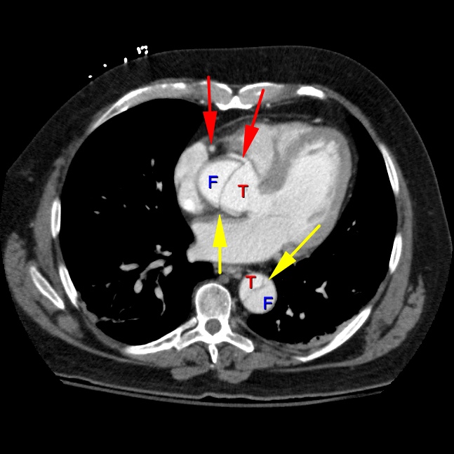
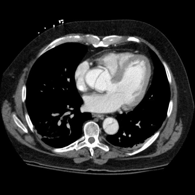
Axial contrast enhanced CT image of the chest at the level of the coronary arteries (red arrows) demonstrates a dissection flap (yellow arrow) with a true lumen (T) and false lumen (F). Since the aortic dissection begins before the left subclavian artery, it is considered a Stanford Type A.


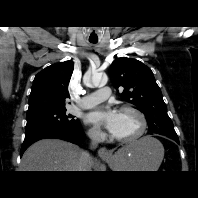
Coronal contrast enhanced CT image of the chest at the level of the aortic arch demonstrates a dissection flap with a true lumen ( T and red highlight) and a false lumen (F). Note the false lumen extending into the brachiocephalic artery. View the next key finding.


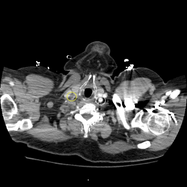
Axial contrast enhanced CT image of the chest at the level of the carotid bulbs. There is near total occlusion of the right common carotid artery (yellow arrow) secondary to the dissection flap. Compare the abnormal right common carotid artery (yellow circle) with the normal left side (white circle). This finding demonstrates a reason why Stanford A type of aortic dissections are surgical emergencies. If the dissection begins distal to the takeoff of the left subclavian artery, it is considered a type B and can be treated more conservatively.

Additional Findings


Return Continue

© 2012 Must See Radiology

MSR

Case #31

Diagnosis

Aortic Dissection
Stanford A

Discussion

This patient presented to an outside hospital with vague abdominal pains. A contrast enhanced CT demonstrated an aortic dissection with the flap involving a portion of the proximal SMA. The scan included a large portion of the chest and it was suggested by the radiologist that the dissection appeared to be involving the aortic root, although not well visualized. The patient was transferred to our hospital for emergency vascular surgery. After speaking with the surgeon regarding the case, he revealed the patient's sister had Marfan's disease, a condition associated with a higher risk for aortic dissection.

Stanford Type A:

  • Involves AT LEAST the ascending aorta
  • Ascending aorta includes just distal to the subclavian artery
  • approx. 60% of all aortic dissections
  • Requires surgery because:
    1. Pericardial tamponade
    2. Coronary artery occlusion
    3. Aortic insufficiency
  • Also classified as either DeBakey type I or type II

Stanford Type B:

  • Inolves only the descending aorta
  • Starring point is distal to subclavian artery
  • approx. 40% of all aortic dissections
  • Does not require surgery unless:
    1. Ischemic extremity
    2. Mesenteric ischemia
    3. Rupture
    4. Aneurysm enlargement of false lumen
  • Also classified as DeBakey Type III

References / Resources

Additional Information:

  1. Dahnert*: 627-630
  2. Primer*: 477-480
  3. Related Article:

    Fisher, ER "Acute Aortic Dissection: Typical and Atypical Imaging Features" Radiographics 1994: 14, 1263-1271.

    McMahon, MA "Multidetector CT of Aortic Dissection: A Pictorial Review" Radiographics 2010: 30, 445-460.


*Dahnert,Wolfgang. "Radiology Review Manual" 7th ed. 2011 LWW.
*Weissleder, Ralph. "Primer of Diagnostic Imaging" 5th ed. 2011 Elsevier.


Return Continue

© 2012 Must See Radiology

MSR

Case #31

Quiz Summary

Not available at this time.

Rate this Case

Rating not available at this time.

Feedback

Any feedback regarding this case can be emailed to Tony@mustseeradiology.com


Thank you for trying Must See Radiology!

Browse All Cases Next Chest Case Next # Case


© 2012 Must See Radiology