
Case #28
-
History: 72 year old with severe mid-abdominal pain. No recent prior surgery.
© 2012 Must See Radiology
History: 72 year old with severe mid-abdominal pain. No recent prior surgery.
© 2012 Must See Radiology

Axial abdominal CT image at the level of the kidneys

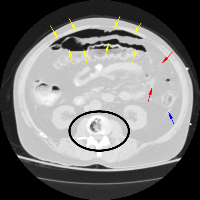
Axial abdominal CT images with body W/L (top image) and lung W/L (bottom image.) Findings include a large collection of free air (pneumoperitoneum), yellow arrows. Lung windows sometimes help decipher fat density from air density on abdominal CT. There is pericolonic fat stranding (red arrow) and peritoneal thickening (blue arrow). Small collections of free fluid also seen throughout, not captured in above image. Incidental finding of vacuum joint phenomenon (secondary to advanced disc disease) through the lumbar spine (black circle)
© 2012 Must See Radiology
Ruptured Transverse Colon
Pneumoperitoneum
This patient presented with spontaneous pneumoperitoneum (i.e. not secondary to iatrogenic trauma). Pneumoperitoneum is most often caused by surgical manipulation within the abdomen. However, in cases that present with no recent surgical history, the finding becomes critical. Finding unexpected free intraperitonal air on a CT scan usually requires immediate surgical intervention.
Differential Diagnosis of Pneumoperitoneum (not related to surgery):
1. Perforated gastric or duodenal ulcer
2. Cecal perforation / Colonic obstruction
3. Pneumotosis Coli
4. Air through the genital tract in females
5. Perforated distal bowel / Diverticulitis, IBD, tumor
This patient underwent emergent laparotomy. a 3 cm rent was found in the transverse colon. The actual tear was not definitively identified on the CT (I think I can see it on the sagittals, but not for sure). There was approximately 250 cc of fecal material also present within the peritoneal cavity. The distal bowel was palpated intraoperatively with no definite obstructing mass identified. No definite cause was identified during the operation for the patient's obstruction and rupture. The colon was repaired and the surgeon performed peritoneal lavage. The patient improved and left the hospital.
Also, it should be noted that in this case, the most amount of free air was around the transverse colon. Keep this in mind when you find free air, as its location does matter.
Additional Information:
Zissin R"Abdominal CT Findings in small bowel perforation." BJR 2009: 82, 162-171.
© 2012 Must See Radiology
Not available at this time.
Rating not available at this time.
Any feedback regarding this case can be emailed to Tony@mustseeradiology.com
Thank you for trying Must See Radiology!
© 2012 Must See Radiology