
Case #29
-
History: Sharp right lower quadrant pain for the last 1.5 days, worsening with worsening pain in the pelvis.
© 2012 Must See Radiology
History: Sharp right lower quadrant pain for the last 1.5 days, worsening with worsening pain in the pelvis.
© 2012 Must See Radiology
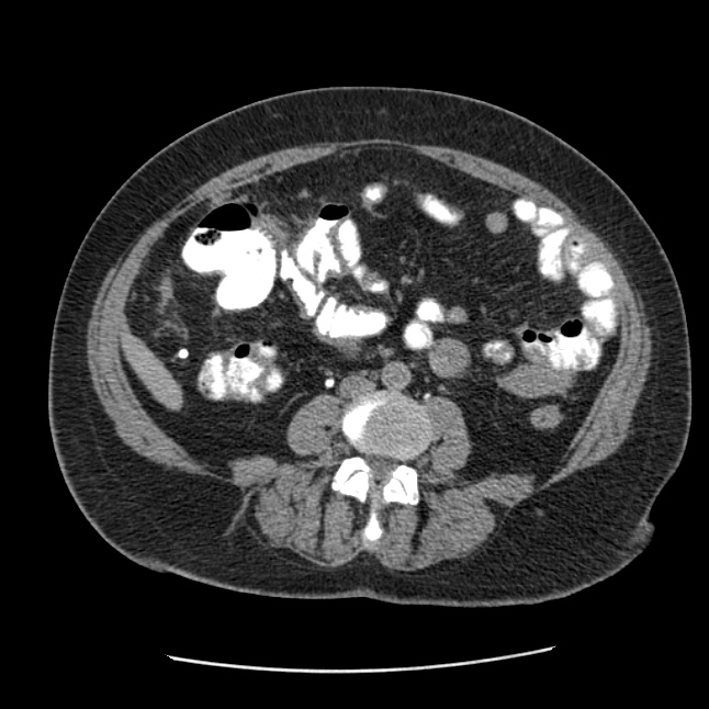
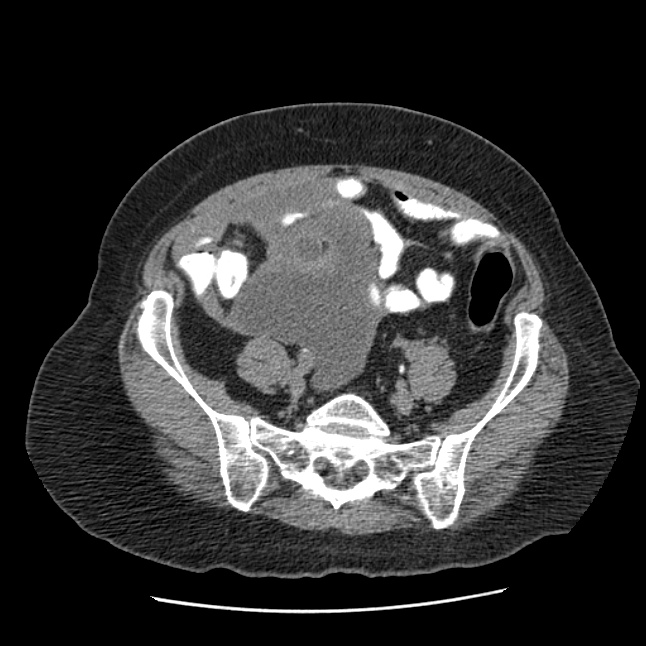
Axial CT abdomen images in the lower abdomen and pelvis
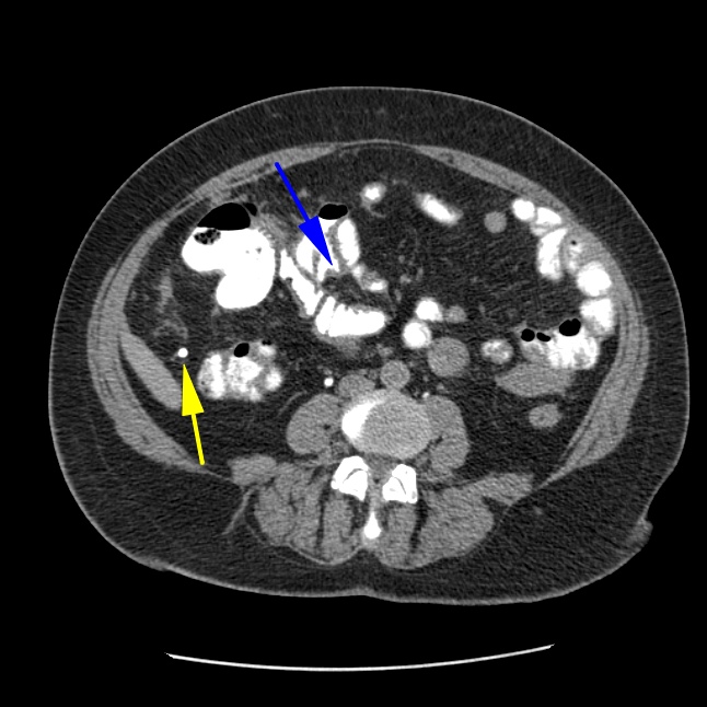

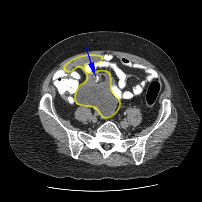
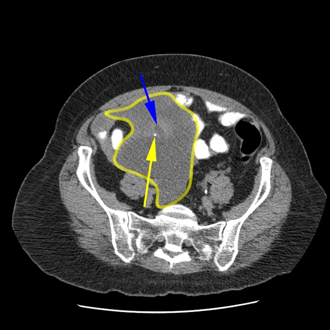
Serial CT images through the lower abdomen demonstrate the appendix (blue arrows) that ruptures into a large fluid collection (yellow outline). Multiple round calcifications are identified in the peritoneum, which may represent appendicoliths (yellow arrow).
© 2012 Must See Radiology
Ruptured Appendicitis
Rupture of the appendix is a complication of acute appendicitis detected too late. This patient presented after rupture had occurred. The patient was treated with antibiotics and the patient underwent percutaneous drainage and catheterization of the fluid collection in the low abdomen.
Additional Information:
Leite NP. "CT Evaluation of Appendicitis and Its Complications: Imaging Techniques and Key Diagnostic Findings." August 2005 AJR, 185, 406-417.
Horton KM. "CT Evaluation of the Colon: Inflammatory Disease." March 2000 Radiographics: 20, 399-418.
© 2012 Must See Radiology
Not available at this time.
Rating not available at this time.
Any feedback regarding this case can be emailed to Tony@mustseeradiology.com
Thank you for trying Must See Radiology!
© 2012 Must See Radiology