
Case #17
-
History: 56 year old male with sharp epigastric abdominal pain for 1 day. Chronic epigastric pain for months, but not as severe as today.
© 2012 Must See Radiology
History: 56 year old male with sharp epigastric abdominal pain for 1 day. Chronic epigastric pain for months, but not as severe as today.
© 2012 Must See Radiology
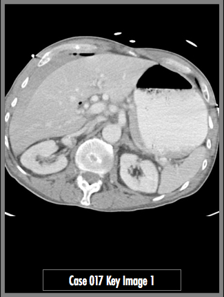
Contrast Enhanced CT of the upper abdomen.
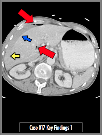
Pneumoperitoneum (red arrows). Free intraperitoneal fluid also seen along the liver. Note the change in density of the fluid from the blue arrow to the yellow arrow. The higher contrast fluid may represent layering hemoperitoneum.

## ADDL IMAGES ##
© 2012 Must See Radiology
Pneumoperitoneum from Ruptured Duodenal Ulcer
No definite ulceration was identified on the original radiology scan. Given the acute abdomen clinical presentation and pneumoperitoneum finding, the patient underwent emergency laparotomy. A duodenal ulcer was found and repaired.
The most common reason for pneumoperitoneum is post-laparotomy or other abdominal surgical procedure. In the patient without a history of surgery, the most likely causes include: peptic perforation, ischemia, and bowel obstruction. One of the most common places for small collections of free intraperitoneal air is in the right upper quadrant. The location of free air is important, especially if it is small. The location of air can clue you into the location of the perforation.
Additional Information:
Zissin R"Abdominal CT Findings in small bowel perforation." BJR 2009: 82, 162-171.
© 2012 Must See Radiology
Not available at this time.
Rating not available at this time.
Any feedback regarding this case can be emailed to Tony@mustseeradiology.com
Thank you for trying Must See Radiology!
© 2012 Must See Radiology