
Case #19
-
History: Right lower quadrant pain, nausea and vomiting. Symptoms much worse today
© 2012 Must See Radiology
History: Right lower quadrant pain, nausea and vomiting. Symptoms much worse today
© 2012 Must See Radiology
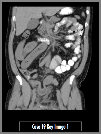
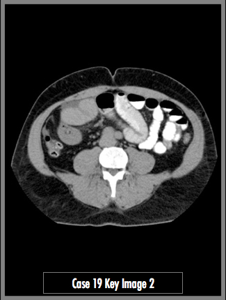
Contrast enhanced CT images of the abdomen and pelvis in the coronal and axial planes
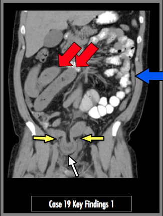
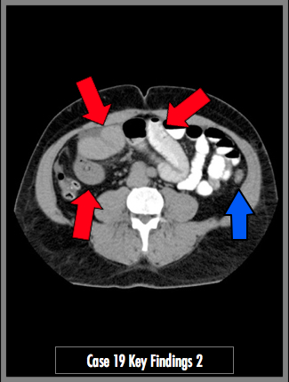
CT abdomen and pelvis axial and coronal images demonstrate multiple loops of dilated small bowel, >2.5 cm diameter (red arrows). There is a loop of distal ileum herniated through the right inguinal canal (yellow arrows). The incarcerated bowel demonstrates bowel wall thickening and surrounding edema (white arrow). The small bowel and colon distal to the obstruction is nearly entirely collapsed (blue arrow).
## ADDL IMAGES ##
© 2012 Must See Radiology
Small Bowel Obstruction / Right Inguinal Hernia
This patient has a loop of distal small bowel that has become invaginated in a right inguinal hernia. The loop of bowel is not reducible and the patient underwent reparative surgery.
The loop of bowel within the hernia demonstrates wall enhancement and surrounding fluid. Note the large proximal loops of small bowel. The diameter of these loops measured up to 3.5 cm. There is a short segment of ileum after the obstruction that is decompressed prior to the ileocecal valve. The entire colon is decompressed. It is important to realize that the severity of bowel obstruction is measured by the degree of decompression in the distal loops, not the width of the proximal loops. To diagnose a small bowel obstruction, there should be at least a few loops of bowel with diameter greater than 2.5 cm. The appearance of the scout radiograph prior to CT exam (available in case menu) is diagnostic for a small bowel obstruction.
Additional Information:
Silva, AC "Small Bowel Obstruction: What to Look For." Radiographics 2009: 29, 423-439.
Boudiaf, M. "CT Evaluation of Small Bowel Obstruction." Radiographics 2001: 21, 613-624.
© 2012 Must See Radiology
Not available at this time.
Rating not available at this time.
Any feedback regarding this case can be emailed to Tony@mustseeradiology.com
Thank you for trying Must See Radiology!
© 2012 Must See Radiology