
Case #20
-
History: 53 year old female with acute substernal chest pain. Evaluation for pulmonary embolism. CT per PE protocol.
© 2012 Must See Radiology
History: 53 year old female with acute substernal chest pain. Evaluation for pulmonary embolism. CT per PE protocol.
© 2012 Must See Radiology
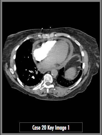
Axial contrast enhanced CT (pulmonary artery CTA, PE protocol)
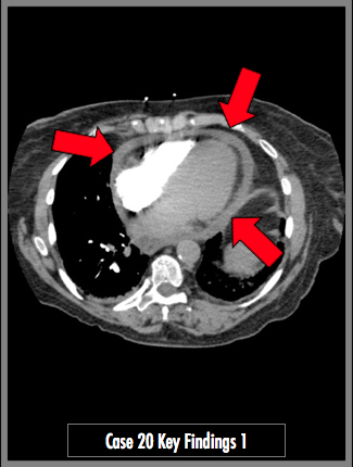
Thickening of the pericardial sac (red arrows). No evidence for thrombus in the pulmonary arteries.
## ADDL IMAGES ##
© 2012 Must See Radiology
Pericardial Effusion
Pericardial Effusion most often presents with dyspnea and fatigue.
Etiology may be from many different causes, including serous fluid, blood, lymph or fibrin. This case demonstrated water density. No signs for cardiac tamponade present in this case.
Cardiac Tamponade may be present in ~50% of cases of pericardial effusion. Cardiac tamponade is a life-threatening condition that results from slow or rapid heart compression secondary to accumulation of fluid, pus, blood, gas, or tissue within the pericardial cavity. CT findings associated with cardiac tamponade include:
- pericardial effusion (usually large) with distention of the SVC & IVC
- reflux of contrast material into the azygos vein and inferior vena cava
- deformity and compression of the cardiac chambers
- angulation or bowing of the interventricular septum.
Additional Information:
Restrepo CS. "Imaging findings in cardiac tamponade with emphasis on CT." RadioGraphics 2007, 27(6): 1595 - 1610
© 2012 Must See Radiology
Not available at this time.
Rating not available at this time.
Any feedback regarding this case can be emailed to Tony@mustseeradiology.com
Thank you for trying Must See Radiology!
© 2012 Must See Radiology