
Case #21
-
History: Child with fever and upper abdominal pain for past few days. History of asthma. PA and lateral chest x-ray and CT abdomen ordered from E.D.
© 2012 Must See Radiology
History: Child with fever and upper abdominal pain for past few days. History of asthma. PA and lateral chest x-ray and CT abdomen ordered from E.D.
© 2012 Must See Radiology
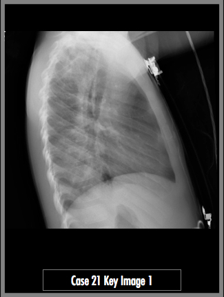
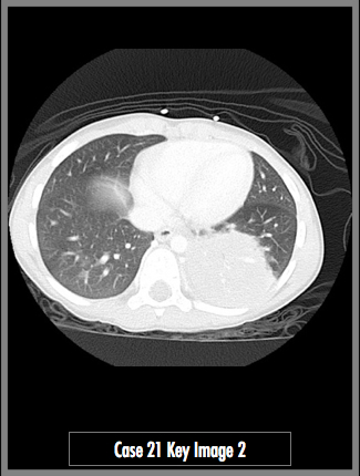
Lateral Radiograph of the chest and Axial CT image through the lower chest
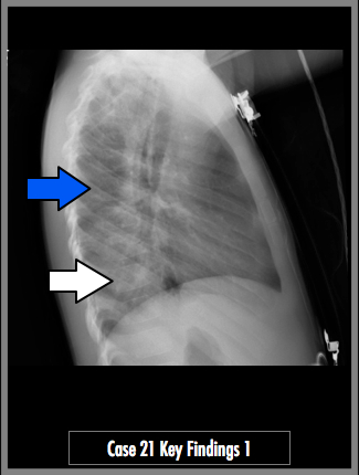
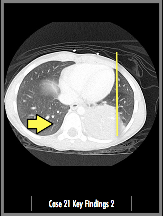
The lateral radiograph of the chest demonstrates an opacity over the lower thoracic vertebral bodies. Observe the difference between the mid-thorax (blue arrow) and the lower thorax (white arrow). The vertebral bodies should appear more lucent from mid to lower thorax. The axial CT image demonstrates an airspace consolidation in the left lower lobe. The consolidation is entirely behind the heart (yellow line) in the AP projection. Given its location, the consolidation is hidden on the frontal radiograph. Imagine the lateral radiograph plane (yellow arrow). Note the amount of overlap between the posterior lung and the vertebral bodies.
## ADDL IMAGES ##
© 2012 Must See Radiology
Left Lower Lobe Pneumonia (Retrocardiac)
The radiographs in this case may appear normal to a radiologist in training, especially on this screen! The lateral radiograph demonstrates an opacity over the posterior lower lobes. In a normal patient, the vertebral bodies should become more radiolucent from the mid thoracic spine to the lower thoracic spine. In this patient, the vertebral bodies appear as the same density or more dense as they approach the diaphragm. When dense appearing lower vertebral bodies are present, review the PA radiograph closer for any airspace consolidation. Since the right lower lobe appears clear, the consolidation is located behind the heart in the left lower lobe.
This child presented to the ER with acute abdominal pain and a history of asthma. Based on the order of a chest radiograph and an abdominal CT, the diagnosis of pneumonia wasn't high on the ER physician's list. However, the suggested differential diagnosis of acute abdomen in children, according to Dahnert, is:
1. Intussusception
2. Appendicitis
3. Obstruction (previous surgery, hernia)
4. Acute gastroenteritis
5. BASILAR PNEUMONIA
Some of my favorite calls to make are on cases that are ordered for different reasons. This is where radiologists can really help a patient and an ordering physician who might be fishing for answers!
Additional Information:
Bramson, RT. "Interpretation of Chest Radiographs in Infants with Cough and Fever." Radiology July 2005: 236, 22-29.
© 2012 Must See Radiology
Not available at this time.
Rating not available at this time.
Any feedback regarding this case can be emailed to Tony@mustseeradiology.com
Thank you for trying Must See Radiology!
© 2012 Must See Radiology