
Case #12
-
History: 13 year old fell off his bike and landed on his left elbow. Pain and decreased range of motion in the left arm.
© 2012 Must See Radiology
History: 13 year old fell off his bike and landed on his left elbow. Pain and decreased range of motion in the left arm.
© 2012 Must See Radiology
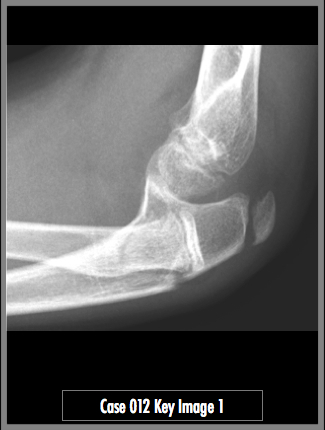
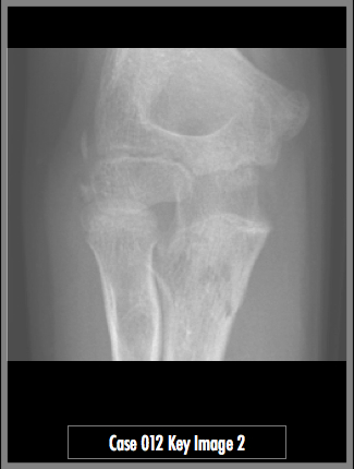
Lateral and AP radiographs of the left elbow.
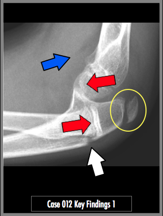
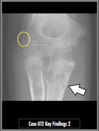
Radiographs of the left elbow demonstrate a fracture along the dorsal aspect of the ulna (white arrow). There is dislocation of the radial head posteriorly from the capitellum (The red arrow tips should point at each other). There is an elbow joint effusion with an anterior humeral fat pad sail sign (blue arrow). Normal medial / "Internal" humeral epicondyle and olecranon ossification centers are present and should not be confused as fractures (yellow circles)
## ADDL IMAGES ##
© 2012 Must See Radiology
Monteggia Fracture (Ulnar fracture / Radial head dislocation)
The pediatric elbow can be a difficult study to interpret for the non-musculoskeletal or pediatric radiologist. Depending on the age of the patient, different ossification centers are present and then are fused or nonfused. In this case, there is a fracture just distal to a non-fused olecranon process.
CRITOE:
The pneumonic CRITOE may help you remember all the ossification centers. Capitellum, Radial head, Internal humeral condyle, Trochlea, Olecranon process, External humeral condyle.
MONTEGGIA:
This case is considered a Monteggia fracture because the combination of ulnar shaft fracture and radial head dislocation is present. There are 4 types of Monteggia type fractures, according to Bado classification:
Bado type 1: 'classic Monteggia' anterior angle ulnar fracture with anterior dislocation of radial head
Bado type 2: 'reverse Monteggia' dorsal angle ulnar fracture with posterior dislocation of the radia head
Bado type 3: ulnar metaphyseal fracture, anterior/anteromedial dislocation of radial head
Bado type 4: proximal 1/3 radius and ulna fracture, anterior displacement of radial head.
Additional Information:
John, SD. "Improving Detection of Pediatric Elbow Fractures by Understanding their Mechanics." Radiographics Nov 1996 16: 1443-1460.
Iyer, RS. "Pediatric Bone Imaging: Imaging Elbow Trauma in Children- A Review of Acute and Chronic Injuries." AJR May 2012 198: 1053-1068.
© 2012 Must See Radiology
Not available at this time.
Rating not available at this time.
Any feedback regarding this case can be emailed to Tony@mustseeradiology.com
Thank you for trying Must See Radiology!
© 2012 Must See Radiology