
Case #16
-
History: 7 month old child fell from sitting, hit side of head on ground. Bump on left side of head. No child abuse suspected.
© 2012 Must See Radiology
History: 7 month old child fell from sitting, hit side of head on ground. Bump on left side of head. No child abuse suspected.
© 2012 Must See Radiology
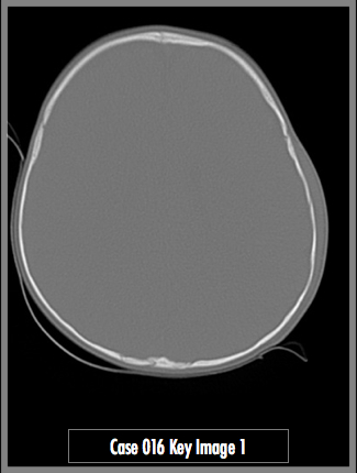
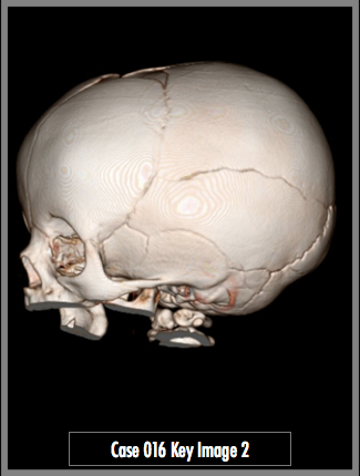
Axial noncontrast head CT (bone window/level) and 3D reconstruction from CT
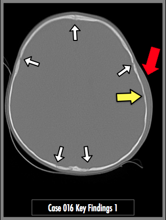
The axial axial head CT demonstrates a mild amount of scalp swelling along the left side of the head. There is a small break in the cortex identified by the yellow arrow that largely resembles the normal unfused suture lines (white arrows).
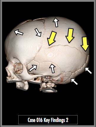
The 3D reconstruction demonstrates a large horizontal fracture along the left parietal bone (yellow arrows). It extends from coronal suture (anterior) to the lambdoid suture (posterior), just above the squamous suture (white arrows).
## ADDL IMAGES ##
© 2012 Must See Radiology
Pediatric Skull Fracture
At my training institution, the majority of residents and attendings use 3D reconstruction on all pediatric head CTs with any trauma-type history. As you can see, finding a fracture can be difficult with the unfused head sutures. When the fracture line is parallel with the axial head CT slice, it is almost impossible. Use of an anatomy reference to identify normal sutures is essential for the radiologist in training (i.e. Netter's Atlas of Human Anatomy 3rd ed, Plate 11). In pediatric and adult head CTs, you can use scalp hematomas and/or abrasions to help localize intracranial/cranial findings.
The presence of a fracture of the skull alone without associated clinical abnormalities of the sensorium or the central nervous system is of little significance. The presence of a fracture should not alter the decision for future medical care except in children with a depressed or compound fracture.
Additional Information:
Harwood-Nash, DC. "The Significance of Skull Fractures in Children, A Study of 1,187 Patients." Radiology October 1971, 101: 151-155.
© 2012 Must See Radiology
Not available at this time.
Rating not available at this time.
Any feedback regarding this case can be emailed to Tony@mustseeradiology.com
Thank you for trying Must See Radiology!
© 2012 Must See Radiology