
Case #13
-
History: Right sided acute chest pain, shortness of breath, elevated D-dimer
© 2012 Must See Radiology
History: Right sided acute chest pain, shortness of breath, elevated D-dimer
© 2012 Must See Radiology

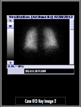
Ventilation-Perfusion Scintigraphy. Perfusion images obtained after administration of Technetium 99-m via IV injection (image 1). Ventilation images obtained after administration of Xenon via inhalation (image 2).
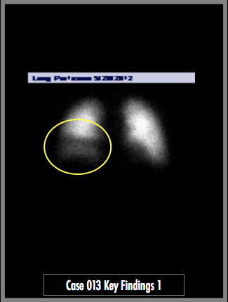
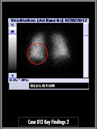
There is a large mismatched perfusion defect in the right middle and lower lobes. No ventilation defect. No pleural effusion or focal airspace consolidation on the chest radiograph. CT interpretation in case #14.
## ADDL IMAGES ##
© 2012 Must See Radiology
High Probability for Pulmonary Embolism
Companion case for this study is case 014, the contrast enhanced CT chest, available in the case menu.
Additional Information:
Sostman, HD. "Acute Pulmonary Embolism: Sens and Spec of Ven-Perf Scintigraphy in PIOPED II Study." Radiology Jan 2008, 246:941-946.
Stein, PD. "Contrast enhanced multidetector spiral CT of the chest and lower extremities in the suspected acute pulmonary embolism: results of the PIOPED II." NEJM 2006, 354:2317-2327.
The PIOPED Investigators. "Value of the ventilation/perfusion scan in acute pulmonary embolism: results of the Pulmonary Embolism Diagonsis(PIOPED)". JAMA 1990, 263: 2753-2759.
Wittram C. "How I do it: CT pulmonary angiography." AJR 2007, 188: 1255-1261.
© 2012 Must See Radiology
Not available at this time.
Rating not available at this time.
Any feedback regarding this case can be emailed to Tony@mustseeradiology.com
Thank you for trying Must See Radiology!
© 2012 Must See Radiology