
Case #22
-
History: 19 year old male with scrotum pain for 2 days.
© 2012 Must See Radiology
History: 19 year old male with scrotum pain for 2 days.
© 2012 Must See Radiology
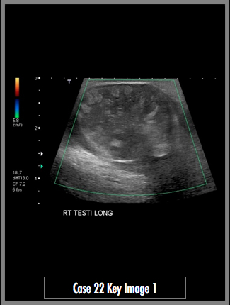
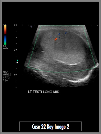
Sonographic images of both testicles in longitudinal plane with a color doppler overlay
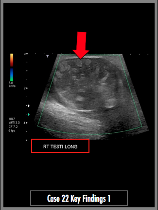
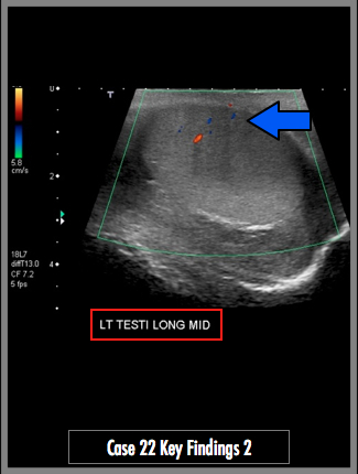
Note the difference in appearance between the left and right testicle (use your labels in red!). The normal left testicle has a homogenous appearance with a few vessels showing flow on color doppler images (blue arrow). The abnormal right testicle has a heterogenous appearance with alternating foci of increased and decreased echoes (red arrow). No internal blood flow is identified in the right testicle.
## ADDL IMAGES ##
© 2012 Must See Radiology
Testicular Torsion (Subacute with Testicular Infarction)
Recognizing acute testicular torsion in patients is important because a delay in treatment may upgrade the patient from a detorsion procedure to an orchiectomy. If the torsion is found within 4 hours, the testicle is usually salvageable. If more than 24 hours pass, the testicle will need to be removed.
- Early findings of torsion include: absent or decreased flow, enlargement, and heterogenous appearance in the affected testicle.
- Late findings include atrophy, reactive hydroceles, and peritesticular inflammation / increased flow.
The patient in this case presented 3 days after the onset of pain. The testicle was infarcted at the time of ultrasound. The alternating echogenic nodularity on ultrasound corresponds with alternating hemorrhagic and infarcted testicle found on surgical pathology.
The surgeon noted that the right testicle was torsed 720 degrees, appeared dark and necrotic. Right orchiectomy was performed. The left testicle appeared normal and left orchipexy was performed.
Additional Information:
Dogra, VS. "Sonography of the Scrotum." Radiology April 2003: 227, 18-36.
© 2012 Must See Radiology
Not available at this time.
Rating not available at this time.
Any feedback regarding this case can be emailed to Tony@mustseeradiology.com
Thank you for trying Must See Radiology!
© 2012 Must See Radiology