
Case #11
-
History: Right Upper Quadrant pain and nausea in an elderly female
© 2012 Must See Radiology
History: Right Upper Quadrant pain and nausea in an elderly female
© 2012 Must See Radiology
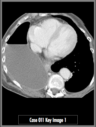
Contrast enhanced CT of the abdomen and pelvis at the level of the lung bases.
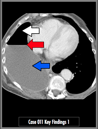
Partial visualization of the lung bases demonstrates a large right pleural effusion (blue arrow) with an air fluid level (red arrow). Pneumothorax (white arrow) confirmed on lung windows (shown below)
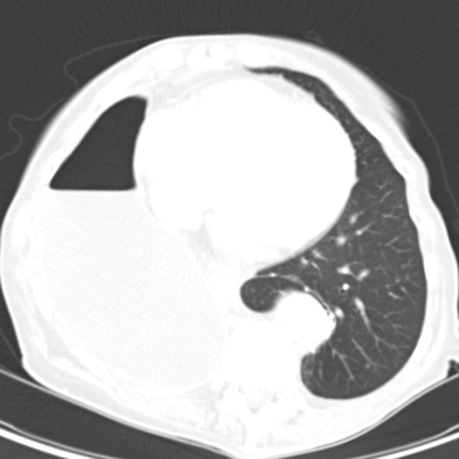
Lung W/L image of the same slice as above. Note the absence of vessels and lung parenchyma in the right pneumothorax, compared to the normal left lung.
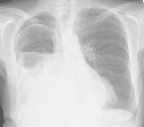
PA radiograph of the chest in the same patient, acquired soon after the CT abdomen. Note the moderate sized pneumothorax in the right apex. There is an air-fluid level in the right hemi-thorax at the level of the right hilum. Near total obscuration of the right heart and right hemi-diaphragm. No findings to suggest tension.
© 2012 Must See Radiology
Hydropneumothorax
This patient presented with abdominal pain and nausea. The patient did not have any respiratory difficulty over her baseline, according to the medical records. The pain may have been referred from the large pleural effusion.
This case represents the incidental findings that you are sure to encounter while on call. Sometimes findings that are not expected are more significant than the presenting problems. This patient likely has chronic lung disease and has recurring pleural effusions. Effusions this large require thoracentesis. Given the pneumothorax, a chest tube is also indicated.
Additional Information:
© 2012 Must See Radiology
Not available at this time.
Rating not available at this time.
Any feedback regarding this case can be emailed to Tony@mustseeradiology.com
Thank you for trying Must See Radiology!
© 2012 Must See Radiology