
Case #10
-
History: Right upper quadrant pain and nausea
© 2012 Must See Radiology
History: Right upper quadrant pain and nausea
© 2012 Must See Radiology
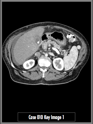
Contrast enhanced CT of the abdomen at the level of the pancreas
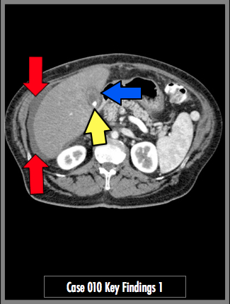
Axial contrast enhanced CT of the upper abdomen demonstrates cholelithiasis (yellow arrow) with gallbladder wall thickening, enhancement and pericholecystic fluid (blue arrow). Perihepatic ascites present (red arrow).
## ADDL IMAGES ##
© 2012 Must See Radiology
Acute Cholecystitis
CT findings of cholecystitis include:
- distended gallbladder
- gallbladder wall thickness >3 mm
- increased gallbladder wall attenuation
- adjacent liver enhancement due to hyperemia
- pericholecystic fat stranding
- pericholecystic fluid
See case #8 and #9 for details regarding the ultrasound and nuclear medicine findings in this case.
Additional Information:
O'Connor, OJ. "Imaging of Cholecystitis." April 2011 AJR 196:4, W367-W374.
© 2012 Must See Radiology
Not available at this time.
Rating not available at this time.
Any feedback regarding this case can be emailed to Tony@mustseeradiology.com
Thank you for trying Must See Radiology!
© 2012 Must See Radiology