
Case #15
-
History: 32 year old female with posterior headaches for 3 days
© 2012 Must See Radiology
History: 32 year old female with posterior headaches for 3 days
© 2012 Must See Radiology
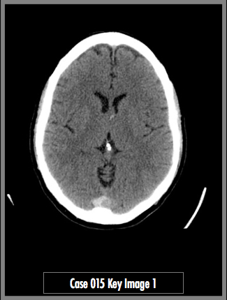
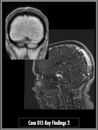
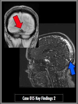
Non-contrast Head CT (image 1) and Coronal Heme Sensitive GRE and Sagittal post contrast T1 MRV (image 2)
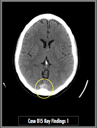
Key Finding 1: Hyper attenuating confluence of venous sinuses with a small filling defect (yellow circle)
Key Finding 2: Susceptibility artifact in the confluence of sinuses on the coronal GRE Heme sensitive sequence (red arrow).
Filling defect in the confluence of sinuses with extension up into the superior sagittal sinus, on the sagittal T1 post contrast MRV sequence (blue arrow).
## ADDL IMAGES ##
© 2012 Must See Radiology
Dural Venous Sinus Thrombosis
Venous Thrombosis can arise from many different causes. Recognizing the subtle radiographic signs of this condition is crucial to proper diagnosis. This case starts with a routine non-contrast head CT that demonstrates hyper attenuation of the sagittal, transverse and straight sinuses. Within the abnormally bright sinuses, the relative hypodense thrombus was identified, which prompted additional imaging with MRI / MRV Brain. Suspicion for venous thrombosis should be high when increased attenuation of the venous system is recognized. However, depending on the stage of disease, sinus thrombosis may appear hyperdense to the surrounding venous blood, as well.
The most common locations for venous sinus thrombosis is the superior sagittal sinus and transverse sinus. If bilateral hemorrhage or infarcts are suspected and no definite arterial distribution is accountable, think of venous sinus thrombosis.
This condition has a high mortality from secondary infarction/hemorrhage.
Additional Information:
Leach JL. "Imaging of cerebral venous thrombosis: current techniques, spectrum of findings, and diagnostic pitfalls." RadioGraphics 2006,26: S19-41.
© 2012 Must See Radiology
Not available at this time.
Rating not available at this time.
Any feedback regarding this case can be emailed to Tony@mustseeradiology.com
Thank you for trying Must See Radiology!
© 2012 Must See Radiology