
Case #8
-
History: Right upper quadrant pain for weeks, worse after meals, nausea, much worse today
© 2012 Must See Radiology
History: Right upper quadrant pain for weeks, worse after meals, nausea, much worse today
© 2012 Must See Radiology


Sonographic images of the right upper quadrant, focused on gallbladder in long and trans projections
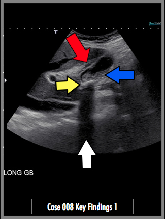
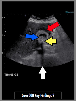
Transabdominal views of gallbladder demonstrate echogenic gallstones (yellow arrow) with posterior clean shadowing (white arrow). There is gallbladder wall thickening, measuring 8 mm (red arrow). Layering tumefactive sludge is also present (blue arrow).
## ADDL IMAGES ##
© 2012 Must See Radiology
Acute Cholecystitis
US evaluation is 81-100% sensitive with >92% negative and positive predictive value (NPV and PPV).
Potential findings include GB wall thickening >3 mm, hazy delineation of GB wall, gallbladder wall lucency or striated wall thickening. GB hydrops (distension with AP diameter >5 cm). Crescent-shaped loculated pericholecystic fluid. Echogenic gallstones with or without impacted stones in the GB neck.
For CT findings in this same patient, please see Case #10
For Nuclear Medicine findings in this same patient, please see Case #9
Additional Information:
Bortoff, GA. "Gallbladder Stone: Imaging and Intervention." May 2000 RadioGraphics: 20, 751-766.
© 2012 Must See Radiology
Not available at this time.
Rating not available at this time.
Any feedback regarding this case can be emailed to Tony@mustseeradiology.com
Thank you for trying Must See Radiology!
© 2012 Must See Radiology