
Case #24
-
History: Patient fell down stairs. Facial swelling and pain.
© 2012 Must See Radiology
History: Patient fell down stairs. Facial swelling and pain.
© 2012 Must See Radiology
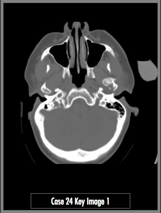
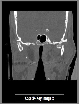
Axial and coronal CT images of the head, through the level of the mandible condyles.
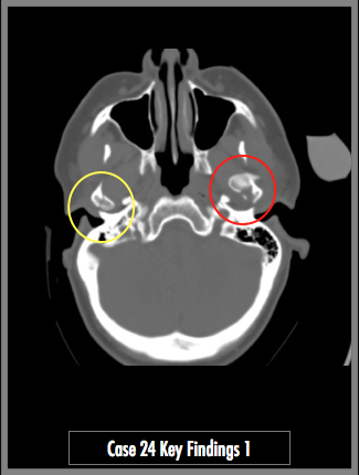
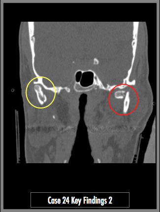
Nondisplaced fracture through the right mandibular condyle (yellow circle). This bony element remains within its joint space. On the left, the mandibular condyle is fractured and displaced medially and anteriorly from its joint space (red circle).

KEY_FINDING_3_TEXT

KEY_FINDING_4_TEXT

KEY_FINDING_5_TEXT

KEY_FINDING_6_TEXT

KEY_FINDING_7_TEXT
© 2012 Must See Radiology
Bilateral mandibular condyle fractures
It is estimated that 30% of mandible fractures occur at the condyle. Given the unique geometry of the mandible and temporomandibular joints (TMJs), these fractures can result in marked pain, dysfunction, and deformity if not recognized and treated appropriately. These fractures may be associated with other injuries that alone have significant morbidity. Examples of such injuries include but are not limited to facial nerve injuries, C-spine injuries, displacement of the mandibular condyle into the middle cranial fossa, injuries to the external auditory canal, and occlusion of the internal carotid artery.
Additional Information:
Kim Goldman et al, "Mandibular Condylar and Subcondylar Fractures" eMedicine August 2012: http://emedicine.medscape.com/article/870075-overview#a1.
© 2012 Must See Radiology
Not available at this time.
Rating not available at this time.
Any feedback regarding this case can be emailed to Tony@mustseeradiology.com
Thank you for trying Must See Radiology!
© 2012 Must See Radiology