
Case #5
-
History: Right flank pain. Sharp, past 1 day. History of left sided kidney stones.
© 2012 Must See Radiology
History: Right flank pain. Sharp, past 1 day. History of left sided kidney stones.
© 2012 Must See Radiology
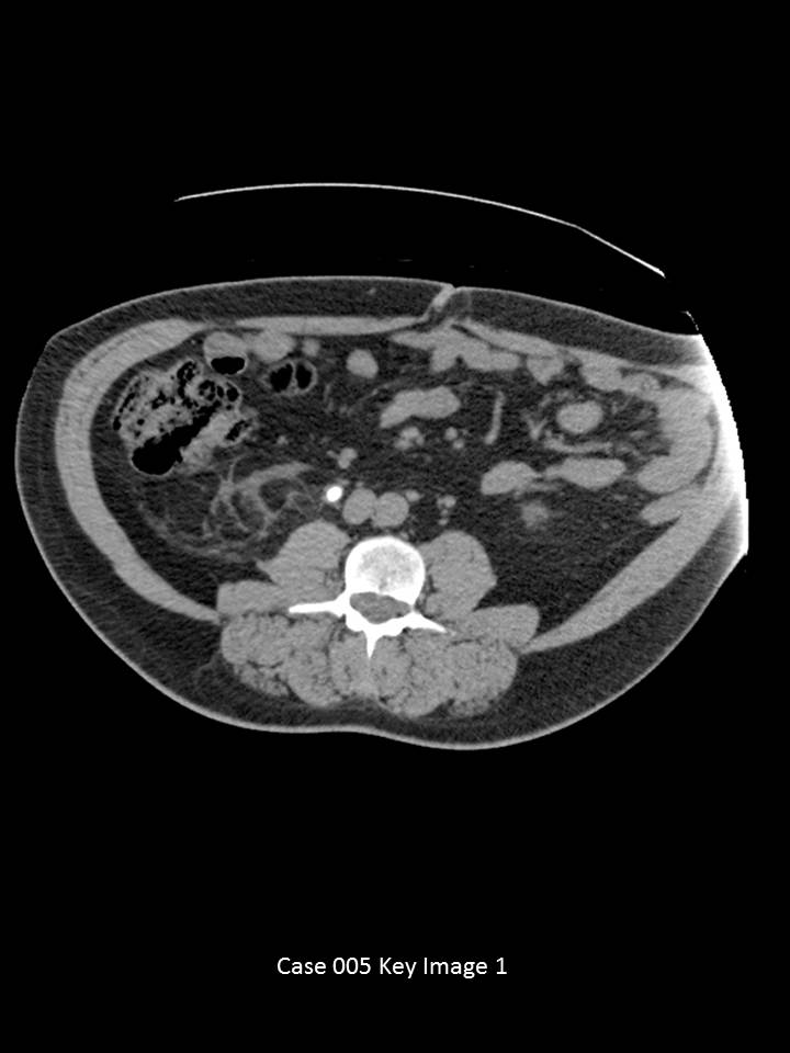
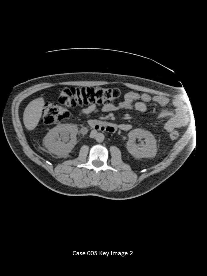
CT abdomen and pelvis without oral or IV contrast through the kidneys and proximal ureters.

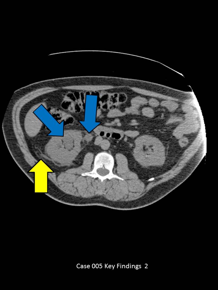
Axial images through the mid abdomen demonstrate a large right mid-ureter calculus measuring 7 mm with a soft tissue rim sign(red arrow). (Soft tissue rim sign is circumferential wall thickening around a stone, which helps differentiate a urinary tract stone from a phlebolith). There is associated right side proximal hydroureter, ureterectasis and pelvicaliectasis (blue arrows). Perinephric fat stranding present on the asymmetrically worse on the right (yellow arrow)
© 2012 Must See Radiology
Nephrolithiasis with right ureter obstruction
This case is an example of high grade ureteral obstruction. The pain is not from the ureter calculus itself, but rather the secondary changes in the proximal urinary system: hydroureter/ureterectasis and hydronephrosis/caliectasis.
When describing obstructive stones, include the size of the stone, location of the stone, presence of proximal urinary tract dilatation and other associated complications. This patient has a history of obstructive nephrolithiasis on the left. It is important to note the other nonobstructing calculi within the renal collecting systems to help the clinician know the stone burden. This patient had a prior CT within the past year that demonstrated the current obstructing calcus as a nonobstructing stone in the lower pelvis of the right kidney.
Prognosis:
Stones <4 mm pass spontaneously in 90%,
Stones 4-7 mm pass in 50%
Stones >8 mm rarely pass spontaneously
Stones <24 mm may respond to extracorporeal shockwave lithotripsy (ESWL)
Stones >25 mm require percutaneous removal
Additional Information:
Tamm, EP. "Evaluation of the Patient with Flank Pain and Possible Ureteral Calculus." August 2003 Radiology: 228, 319-329.
© 2012 Must See Radiology
Not available at this time.
Rating not available at this time.
Any feedback regarding this case can be emailed to Tony@mustseeradiology.com
Thank you for trying Must See Radiology!
© 2012 Must See Radiology