
Case #3
-
History:Acute sharp right flank pain. No history of renal stones. Noncontrast CT Abdomen ordered.
© 2012 Must See Radiology
History:Acute sharp right flank pain. No history of renal stones. Noncontrast CT Abdomen ordered.
© 2012 Must See Radiology
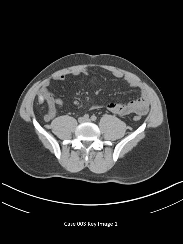
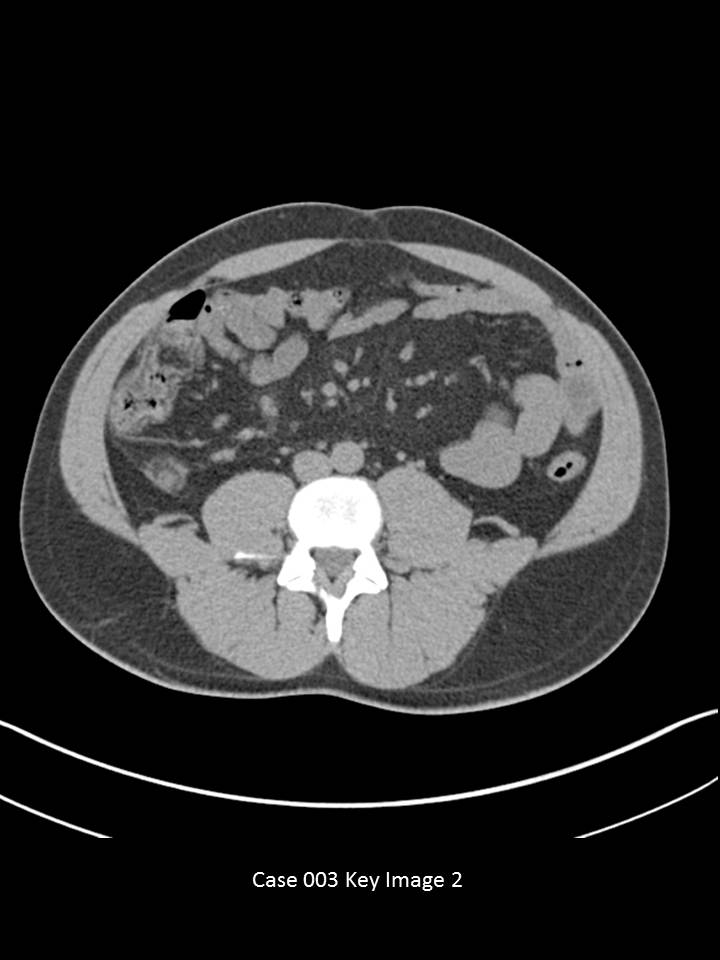
CT Abdomen and Pelvis without IV or oral contrast
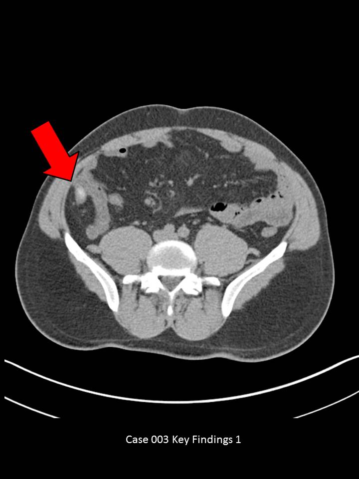
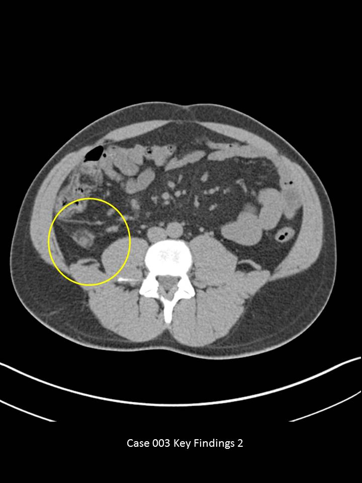
Axial images through the lower abdomen demonstrate an appendicolith (red arrow) in a fluid filled appendix. There is periappendiceal fat stranding (yellow circle). The appendix is significantly dilated, resembling the caliber of adjacent small bowel. No abscess formation, no free intraperitoneal air or significant fluid. No evidence for urolithiasis.
© 2012 Must See Radiology
Acute Appendicitis
This patient presented with vague right flank pain symptoms that started earlier in the day. Findings on CT represent a mild acute presentation of appendicitis. The patient underwent surgery within the day and an inflamed appendix was removed.
The appendix should always be surveyed in the acute abdominal pain setting.
Find the appendix: Locate the ileocecal junction and follow the cecum posteromedially ~1-2 cm. You will find the most difficult appendices to find are the ones that are in patients with RLQ pain! Use your coronal and sagittal images, if needed.
Findings suggestive of acute appendicitis on CT include peri-appendiceal stranding /inflammation, circumferential wall thickening, fluid filled appearance, appendicoliths, apical cecal inflammation/thickening, and/or free peritoneal fluid. Dilatation of the appendix alone is not a strong predictor. Also, gas may be present in a normal appendix or necrotizing appendicitis. Use all of your other clues to decide if the appendix is acutely inflamed.
Complications: Perforation, Periappendiceal Abscess, Peritonitis, Bowel Obstruction, Gangrenous Appendicitis, Septic Seeding of Mesenteric Vessels
Additional Information:
Leite NP. "CT Evaluation of Appendicitis and Its Complications: Imaging Techniques and Key Diagnostic Findings." August 2005 AJR, 185, 406-417.
Horton KM. "CT Evaluation of the Colon: Inflammatory Disease." March 2000 Radiographics: 20, 399-418.
© 2012 Must See Radiology
Not available at this time.
Rating not available at this time.
Any feedback regarding this case can be emailed to Tony@mustseeradiology.com
Thank you for trying Must See Radiology!
© 2012 Must See Radiology