Key Findings
Key Findings
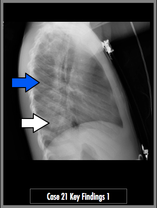
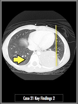
The lateral radiograph of the chest demonstrates an opacity over the lower thoracic vertebral bodies. Observe the difference between the mid-thorax (blue arrow) and the lower thorax (white arrow). The vertebral bodies should appear more lucent from mid to lower thorax. The axial CT image demonstrates an airspace consolidation in the left lower lobe. It is entirely behind the heart (yellow line) in the AP projection, which hides in on the frontal radiograph. Imagine the lateral radiograph plane (yellow arrow). Note the amount of overlap between the posterior lung and the vertebral bodies.