Key Findings
Key Findings
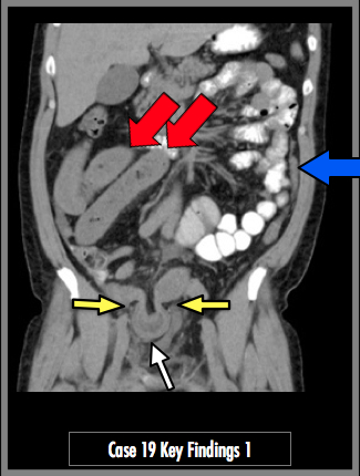
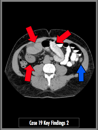
CT abdomen and pelvis axial and coronal images demonstrate multiple loops of dilated small bowel, >2.5 cm diameter (red arrows). There is a loop of distal ileum herniated through the right inguinal canal (yellow arrows). The incarcerated bowel demonstrates bowel wall thickening and surrounding edema (white arrow). The small bowel and colon distal to the obstruction is nearly entirely collapsed (blue arrow).