Key Findings
Key Findings
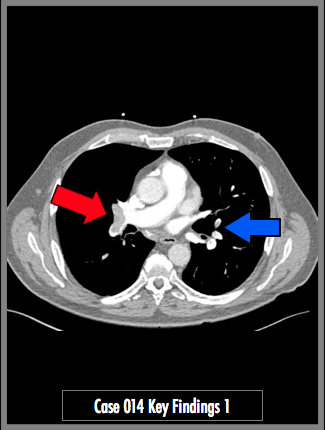
Contrast enhanced CT of the chest in axial and coronal MIP views. Pulmonary arteries are opacified well (>300 HU in the main pulmonary artery). Images demonstrate low density filling defect in the right pulmonary artery branches to the middle and lower lobes, "saddling" both branches (red arrows). Additional small segmental filling defects in the left lower lobe pulmonary artery branches.