Key Findings
Key Findings
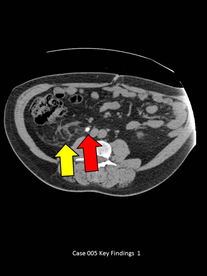
Axial images through the mid abdomen demonstrate a large right mid-ureter calculus measuring 7 mm with a soft tissue rim sign(red arrow). (Soft tissue rim sign is circumferential wall thickening around a stone, which helps differentiate a urinary tract stone from a phlebolith). There is associated right side proximal hydroureter, ureterectasis and pelvicaliectasis (blue arrows). Perinephric fat stranding present on the asymmetrically worse on the right (yellow arrow)